
Fuchs dystrophy: causes and treatment
Fuchs dystrophy is a disease of the cornea that causes blurred vision and progressive visual loss. Know symptoms, diagnosis and treatment.

MAPFRE Health Blog is a leading blog in the world of medicine thanks to the fact that all its contents are written by specialized doctors.
MAPFRE’s years of experience in the sector guarantee us as a source of truthful and practical information, helping you with your questions about the health of the body and mind.
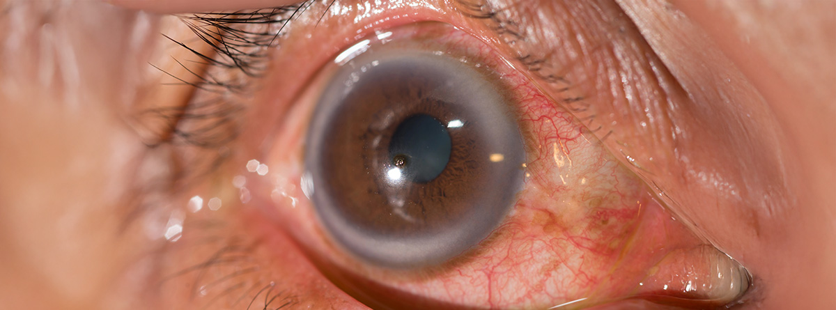
Fuchs dystrophy is a degenerative disease of the cornea that damages the endothelium (the inner layer of cells of the cornea). These cells pump fluid to keep the cornea clear. When its function is lost or fails, fluid accumulates, the cornea swells (edema) and vision deteriorates. One of the first findings is the appearance of guttas (deposits in the innermost layer of the cornea).
Although it is bilateral, can affect both eyes asymmetrically. The disease usually manifests clinically around the age of 50-60, although some subclinical signs may appear between the ages of 30-40.
Causes and risk factors
- Genetics: has a hereditary basis in many cases. Genetic mutations have been identified in several genes, including variants in TCF4 (especially the CTG18.1 expansion), as well as mutations in COL8A2 in early forms or specific families.
- Age: Risk and severity increase with age.
- Sex: It is more common in women than in men.
- Other predisposing factors: Previous cataract surgery can accelerate endothelial loss; oxidative stress, environmental damage, possibly metabolic factors.
What symptoms does it produce?
- Early stage: Blurred vision in the morning, which improves during the day.
- Advanced stage: persistently blurred vision, presence of epithelial blisters (bullae) that can rupture and cause pain. Halos, glare, light sensitivity. Loss of visual acuity (not seen in detail).
How is the diagnosis made?
The diagnosis is made by the ophthalmologist through simple examinations.
- Slit lamp examination: to see the guttae, opacities, corneal edema.
- Endothelial cell count and morphological evaluation by specular microscopy.
- corneal pachymetry: to measure central thickness of the cornea and quantify edema (inflammation).
- Anterior segment optical coherence tomography (corneal OCT) to quantify thickness, edema and classify severity.
In MAPFRE Health Insurance you will find great ophthalmologist specialists who will give you the most precise diagnosis and offer you the most suitable treatment for your ailment.
CALCULATE YOUR PRICE
What are the treatments?
Conservative/Medication
- Hyperosmotic eye drops or ointments (for example, saline solutions with a high concentration of sodium chloride) to reduce edema by removing fluid accumulated in the cornea.
- Lubricantsmoisturizers to relieve discomfort.
Surgical
The only curative treatment is corneal transplant. It can be done in several ways.
- Endothelial keratoplasty: Techniques such as DSAEK, DMEK are preferred in most cases, as only certain layers of the cornea are replaced without the need for a complete transplant, with faster recovery and fewer complications.
- Penetrating keratoplasty: e reserved for cases with extensive damage or when it is not possible to use endothelial techniques. A part of the cornea is removed and the donor cornea is sutured.
The most common complications of surgical treatment are infection, transplant rejection, poor healing and complications with sutures. After surgery, corticosteroid treatment is performed for a long period to prevent rejection.
Research lines
Cellular and regenerative therapies
- Techniques are being developed to cultivate human endothelial cells in vitro and transplant them to the eye without the need for complete grafts.
- Clinical trials in Japan and Europe show that intracameral injection of cultured endothelial cells combined with Rho-kinase (ROCK) inhibitors improves corneal transparency and vision, avoiding keratoplasty in some cases.
Gene therapies
- Aimed at the mutations associated with the disease and thus modulate and stop cellular degeneration.
Experimental drug treatments
- ROCK pathway inhibitors (such as ripasudil and netarsudil) to stimulate regeneration and improve residual endothelial function.
- Some antioxidants and oxidative stress modulators are also being studied.
Less invasive surgical advances
Descemet Stripping Only (DSO) techniques are explored, which consists of removing the Descemet membrane (damaged corneal membrane) without transplant, relying on the migration of healthy cells or after cell grafts.
What you should know…
- Fuchs dystrophy is a degenerative disease of the cornea that affects the endothelium, causing fluid accumulation, edema and progressive loss of vision.
- The diagnosis is made through ophthalmological examination and specific tests such as pachymetry, specular microscopy and OCT, and treatment can be conservative or surgical, with corneal transplant being the only definitive cure.
- Cellular, gene and pharmacological therapies are currently being investigated, as well as less invasive surgical techniques, with the aim of avoiding complete transplantation.